
There exists a variety of living organisms, from which some are visible to the naked eye, and some remain invisible. All these entities are classified into major groups, called prokaryotes and eukaryotes. Today, we are talking about a prokaryotic microorganism, a bacterium, and will see what it looks like under a microscope.
Bacteria are single-celled microscopic bodies that genetically differ from the eukaryotes, and are easily observed through student microscopes. Scientists say these life forms were the first producing bodies on Earth back then, around four billion years ago! We currently live with very deadly bacteria surrounding us, but most of them are pretty useful too.
Nowadays, good bacteria help humans in several ways, whether in growing plants, curing animals, pharmaceutical or industrial processes. Let’s step ahead to learn the basics of this species and consider its shapes under a microscope. Make sure, such observations require high magnification microscopes for microbiologists. This can not work well with all microscopes.
Different Types of Bacteria
These microorganisms have numerous types and specific characteristics, being a diverse living group. Bacteria can reside inside our bodies and in the external environment. They have adjusted themselves in the surroundings for over the years now, including cold temperatures, deep-sea oceans, etc.
- These can be pathogenic or non-pathogenic microscopic bodies, keeping in mind the property of causing disease. Pathogenic bacteria are harmful when they attack a host cell, while the non-pathogenic group proves beneficial.
- Some bacteria can easily grow in environments deficient in oxygen and are referred to as anaerobic bacteria. On the contrary, it’s essential to provide oxygen supply to aerobic bacteria.
- Another classification of bacteria is according to the retention of primary stain. Gram-positive bacteria grow with a thick peptidoglycan layer which allows easy staying of the primary stain. In contrast, a gram-negative bacteria never holds a primary stain on it.
How to Identify a Bacteria Under a Microscope? Step-by-Step Guide
Observing a bacterium cell under any type of microscope follows the same rule as any other specimen. Identifying bacteria, in the beginning, requires culturing it with specific growth factors, and once fully grown, you can keep the slide under the microscope to study it. One of the most important factors to consider in the entire process is to conduct the experiment with high sterilization practices.
Step 1: Culture
Bacteria prevail naturally as single cells, clusters, pairs, bunches, or long chains. If you choose a certain type of bacteria as your specimen, first you have to grow it in a bigger population in an appropriate culture medium.
Types of Culture Mediums
- Nutrient Agar Media: It is the most widely used non-specific medium in which a huge variety of bacterial samples are cultured. They include all aerobic and non-aerobic bacterial organisms. On the basis of oxygen consumption, bacteria are divided on a wide scale.
- Selective Agar Media: Mannitol salt is used as the basic constituent of this medium. It enables the multiplication of certain bacterial species while halting the growth process of all other bacteria introduced in Selective Agar.
- Differential Media: In this medium, different materials are used to identify different bacteria and cultures the one that is required. For example, if you are looking to isolate and cultivate a Streptococcus colony, introducing a Blood Agar Media would be the most appropriate. Streptococci have hemolytic properties and would become prominent if provided such a medium.
Step 2: Prepare a Neat Slide
Once the bacterial incubation period is achieved, it is time to prepare a bacterial smear on a glass slide. Your slide can be viewed under a microscope to unveil the mysteries of bacteria right after this smear is ready.
How to Prepare Bacterial Slide – Step-by-Step Guide
First, you need a marker to mark a certain point on your slide where you will place your bacterial smear.
Secondly, with the help of an inoculation loop, introduce very little water at the spot of smear preparation.
Third, heat the inoculation loop in the blue flame of the Bunsen burner, and let it glow red hot. Now allow the loop to cool down; immediately lift some bacteria from the culture broth with aid of this loop.
Thereafter, mix the bacteria with the water drop on the slide, carefully with the inoculation loop’s end while placing the bacteria.
Lastly, allow this mixture to dry at atmospheric air pressure/room temperature.
Step 3: Time to Stain Your Slide
This straining step is pretty crucial to enable bacterial structures like cell walls, membranes, cytoplasm, and plasmids. Educational institutes and laboratory setups mostly use Methylene blue, Safranin, and Crystal Violet stains for this purpose.
To Stain Your Slides
First, organize them on a staining rack. Introduce the required stain with a dropper, over your bacterial smear, and allow it to dry for one to two minutes.
Next, wash away the excessive staining pigment by passing your slide gently through the slow stream of water, and slightly rinsing it.
Your slide is now ready to be viewed under a microscope.
Adjust it on the stage of your microscope, and make necessary adjustments for a clear specimen’s view, depending upon the focus modifications that your microscope possesses.
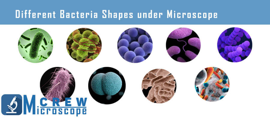
20+ Different Shapes of Bacteria [ Viewed under Microscope ]
We distribute this set of microorganisms into four major classes as per their shapes, so it’s easy to determine them. These include Cocci (spherical-shaped), Bacilli (rod-shaped), Spirilla (spiral-shaped), Vibrio (comma-shaped), and similar others.
Cocci (Spherical-Shaped)

Observing this bacterial shape under a microscope appears to be spherically round, oval, and sometimes bean-shaped. We found multiple sub-types of cocci that were either assorted as two, four, eight, or many. The further sub-divisions are as follows:
- Monococcus: These are the species with only a single cell.
- Diplococcus: As the name suggests, these cocci appear to be in pairs under a microscope, for example, Neisseria lactamica, Moraxella catarrhalis, Neisseria subflava, etc.
- Tetrads: Such bacterial cocci have four cells. Tetrads are distributed among two planes, with common examples as Pediococcus and Aerococcus.
- Streptococcus: These bacteria are seen arranged in a plane, following a unique pattern. Streptococcus has special non-motility properties and is mostly aerobic. Some examples are Streptococcus bovis, Streptococcus mutans, Streptococcus agalactiae, etc.
- Staphylococcus: This sub-class of cocci includes a huge number of bacteria clustered together in irregularity. These are often observed as grapes under the microscope because of the presence of three planes. It covers examples like Staphylococcus haemolyticus, Staphylococcus epidermidis, Staphylococcus aureus, etc.
- Sarcina: The bacterial genus that shows a cuboidal cocci arrangement under microscopic study is called Sarcina. These are usually the anaerobic gram-positive bacteria, so observing them demands primary stain and a proper growth medium. Several examples of these bacteria are present, including Clostridium maximum, Sarcina ventriculi, Micrococcus luteus, etc.

Bacilli (Rod-Shaped)
The bacilli type of bacteria comes out as rod-shaped under the microscope. These specific bacterial profiles can be further subdivided into classes based on the number of bacilli involved. Most rod-shaped bacteria are gram-positive, so easily identified by primarily staining them. Let’s see what the bacilli sub-types look like under a magnifying apparatus:

- Bacillus: It is simply an individual bacterial cell in a rod shape, for example, Salmonella enterica, B. anthracis of anthrax.
- Diplobacilli: If you examine a Diplobacilli, it shows double rods connected to each other, making a group. Some examples are K. rhinoscleromatis, M. bovis, etc.
- Palisades: It gives a fence-like appearance below the microscope after dividing, for instance, C. diphtheria.
- Coccobacilli: These bacteria are a little combination of cocci and bacilli, giving a slightly oval shape, for example, Gardnerella vaginalis.
- Streptobacilli: The Streptobacilli arrange themselves in a chain, being aerobic and gram-negative species. The most common examples of such types of bacteria are Streptobacillus notomytis, Streptobacillus felis, Streptobacillius ratti, etc.
Spirilla (Spiral-Shaped Bacteria)

These bacterial types have a particular spiral shape, often appearing as curves under a microscope.
The Spirilla sometimes shows us curves and sometimes a corkscrew shape.
Most of them are very motile microorganisms. The spiral-shaped microscopic units have a long and flexible sub-class called Spirochete.
Spirochete: Such microorganisms have smooth flagella present, which are very thin and motile. Spirochetes are quite dangerous species, causing many bacterial diseases like Lyme infection and Syphilis.
Vibrio (Comma-Shaped Bacteria)
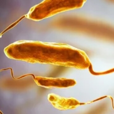
Microscopes detect these organisms in comma shapes that are naturally gram-negative. Vibrio is well-known to cause serious foodborne ailments, being anaerobes. They have a specific characteristic of replicating immediately, thus, spreading diseases.

There are interesting organisms that exist as bacteria protists, and some are even single-celled plants. Yes! We are talking about Planktons. Let’s briefly discuss this organism and see what it looks like under a microscope.
Planktons
Planktons, surprisingly, are a diverse group of organisms that are essential for marine life’s survival. These organisms are numerous and so tiny that one drop of water could have thousands of them, most Planktons are sized 0.05 and 1 mm or 0.002 and 0.04 inches only. Planktons are classified as Phytoplankton and Zooplankton, being plant-like and animal-like in nature, respectively. Most of these microscopic organisms are often less than one inch in size, but some are macroplanktons as well, e.g., jellyfish and sargassums.
Who Do We Identify Planktons?
As discussed earlier, almost all planktons are microscopic whose shapes can be clearly seen under a microscope. These are naturally in several beautiful colors. Some phytoplanktons are irregular-shaped, while most are single-celled and round-shaped. Coming towards zooplankton, many marine organisms swim in the water with large antennae, covered in shell-like structures.
What are Zooplankton?
All those planktons with animal-like characteristics are zooplanktons. They can be as big as five mm in length and as small as even the thousandth part of it, hence, observed under a microscope. Zooplankton exist as unicellular and multicellular organisms. Further, these heterotrophs grab their food and energy from organic compounds and other plants, such as algae.

One interesting feature of zooplanktons is that they drive themselves deep underwater to hide from predators in daylight. On the other side, the same species look towards the water surface to attack phytoplanktons at night.
Marine Planktons
Marine planktons fall under zooplanktons, including marine archaea, bacteria, protozoa, and algae. These organisms spend their lives drifting and floating under seawater or saltwater. Further elaborating on algal groups, all the dinoflagellates, diatoms, green algae, etc., are marine plankton that can be clearly examined under a microscope.
These organisms feed on phytoplanktons and are then eaten by large zooplankton, and the chain goes on. These diverse species survive in the ecosystem, being the backbone of the marine food web.
What are Phytoplanktons?
As we have discussed the role of phytoplanktons before, these microscopic plants are essential to maintain the marine food web. Similar to terrestrial plants, phytoplanktons help convert sunlight into energy for organisms underwater. This photosynthetic procedure takes in carbon dioxide and produces oxygen. It’s interesting to note that almost 50 percent of oxygen underwater is due to the presence of phytoplankton.
Other Bacterial Shapes under Microscope
- Star Shape: The name itself indicates the shape of this prokaryotic bacteria. It appears as a star when observed through a microscope, for example, the Stella species.

- Trichome: These bacteria are born with an extra sheath over them and are observed as vegetative cells. A sulfur-oxidizing bacteria called Thiothrix Nivea is a common example.
- Fusiform: Such bacterial shapes are fused when seen in a microscope and are in spindles, for instance, Fusobacterium necrophorum.
- Pleomorphic: It’s a unique bacterial kind that does not have any defined shape. A pleomorphic bacteria can change its formation as per the external environment, such as Mycoplasma genitalium.
- Sheathed: We can grab these shapes of bacteria from inside water as they’re covered with sheath, for instance, iron bacteria (Leptothrix discophora).
- Rectangular: One can easily determine the structure of this bacteria through its name. These are box-shaped organisms, including halophilic bacteria.
How can I Easily Observe a Bacterium?
Bacteria have a size range, with a diameter usually of 0.2 nano-meter and a length of 2-8 micron meters. You can easily recognize any shape of bacteria under a light or electron microscope, having specific conditions. Students are instructed to grow a bacterium in a definite growth medium and then stain it with the provided dye. Then a biological or compound microscope of high magnification like M150C-I is set to observe details of a bacteria.
Experienced people can conveniently differentiate a bacterium cell from a dust particle, but it takes time for students to identify it instantly.
Leave a Reply