
Light microscopes and electron microscopes are inevitable in scientific research. They provide insight into the structure of different samples like viruses and bacteria. However, electron microscopes are more complex than compound or light microscopes and provide higher magnification. The scanning electron microscope is a type of electron microscope.
Let’s tell you the differences between an electron microscope and scanning electron microscope.
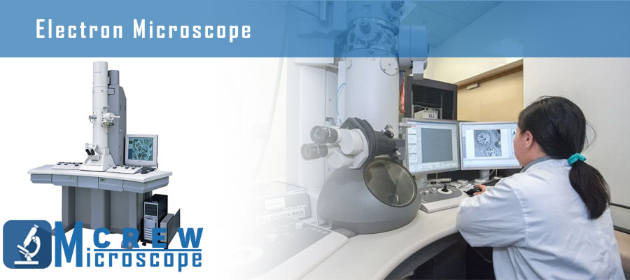
What is an Electron Microscope?
Electron microscopes use electrons produced by a cathode to obtain high-resolution images of non-living biological and non-biological samples. The samples have to be dried to prepare them for electron microscopy. Thus, viewing live germs, cells, or tissues in electron microscopes is not possible. However, researchers know and understand the use of microscope, so they might use electron microscopes to study dead cells, tissues, macromolecules, ribosomes, and mitochondria.
Electron microscopes use short-wavelength electrons as the illumination source to produce high-resolution images. Electron microscopes are sometimes used with other techniques like negative staining, thin sectioning, etc. Electron microscopes have been widely used to inoculate molecules, study compounds, and evaluate diseases.
Electron Microscopy is classified as Transmission Electron Microscopy (TEM) and Scanning Electron Microscopy (SEM). Although, these processes are entirely not the same, but both use an electron source to illuminate the object and produce an image. Transmission electron microscopy helps examine thin specimens by allowing the electrons to pass through them. It resembles the working of a compound microscope in unique ways. However, the detailing obtained by a transmission EM is way higher than a light microscope.
Keep reading to learn everything about scanning electron microscopes in detail.
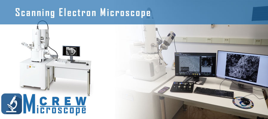
What is a Scanning Electron Microscope?
A scanning electron microscope produces a high-resolution image of the surface of a sample using a focused electron beam. The images created by a scanning electron microscope give information about the topography and composition of the material’s surface.
The electron beam does not pass through the sample in a scanning electron microscope. Instead, it scans the object’s surface under examination to produce a highly magnified image. A scanning electron microscope resolving power is much higher than light and stereo microscopes. It is initially increased by using an electromagnetic lens and further multiplied using a shorter wavelength produced by the electron beam.
The shape, size, and state of the sample is critical to observation in electron microscopes. You need to coat the sample’s surface with a sputter-coater to ensure it conducts the electrons. Coating materials may include chromium, silver, gold, and platinum. Furthermore, sometimes, non-conducting materials like plastic are also used for coating.
The samples should also be dried and fixated before observation using alcohol to maintain the shape and form of the sample.
Now that you know how electron microscopes, scanning electron microscopes, and transmission electron microscopes are related, here are the differences between electron microscopes and scanning electron microscopes.
Differences Between Electron Microscope and Scanning Electron Microscope
Definition
An electron microscope is a type of microscope that uses an electron beam to focus on the sample and produce high-resolution images.
Scanning electron microscope produces images by bombarding the surface of the sample. The electrons do not pass the sample in scanning SEM.
Parts
The electron microscope comprises an electron gun, electromagnetic lenses, a specimen holder, and an image-viewing and recording system.
The scanning electron microscope contains an electron source, anode, scanning coils, condenser, and objective lens.
Working Principle
The electron microscope uses signals from the falling of electrons on the sample to get information about the composition and morphology of the specimen. The electron gun generates electrons that the condenser lenses focus into a thin beam. The specimen is extra thin, and the electrons produce images when they come in contact with the sample. The images show more and less dense regions due to differences in the scattering of electrons. The ocular lens gives the final image after it passes through the objective lens.
The working principle of scanning electron microscopes is almost the same as mentioned above. Nevertheless, different types of electron microscopes use specific working principles. Thus, Scanning EM uses the electron beam to produce a high-resolution image without passing through the sample. The filament emits electrons which are converged into a thin beam. The lenses focus the electron beam and focus the image onto the eyepiece.
Types
Electron microscopes are categorized into scanning electron microscopes and transmission electron microscopes based on their working. A scanning electron microscope produces an image by scanning the surface of the sample. Contrarily, the electron beam transmits through an ultra-thin specimen to produce the observation in electron transmission microscopes.
On the other hand, scanning electron microscopes are not classified into further types. But, they may vary in size and capability. You can choose from TEM solutions, SEM Detectors, and Solutions, Phenom Desktop SEMs, etc.
Functions
Electron microscopes are used to find the morphology of various biological and non-biological samples. They also help in understanding the chemical structure of different molecules and compounds.
Scanning EM can perform several types of analysis, including backscattered electron detection (BSE), cathodoluminescence, electron backscatter diffraction (EBSD), and energy dispersive x-ray spectroscopy (EDS).
Application
Electron microscopes are widely utilized in microbiology research labs besides light microbiology microscopes. They also help in forensics for biopsy samples. Industries often ensure quality control using electron microscopes.
Scanning electron microscopes also help in forensic investigation and medical sciences. Furthermore, their application lies in electronics, geological sciences, and microscopic quality control.
The Bottom Line
Researchers use electron microscopes to study the samples in high magnification and produce high-resolution results. Scanning electron microscopes are a type of electron microscope besides transmission electron microscope. They use an electron beam to illuminate the samples converged by one of the modern types of lenses, electromagnetic lenses. Electron microscopes have vast applications in biology, geology, and electronics.
FAQs
What are two different types of electron microscopes?
Scanning electron microscopes and transmission electron microscopes are the two types of electron microscopes. Sometimes, the TEM and SEM are combined in one instrument known as the scanning transmission electron microscope (STEM).
What is the main difference between SEM and optical microscopy?
Optical microscopes are easier and quicker than scanning electron microscopes. Scanning electron microscopes provide a highly magnified image. Yet, the samples take more time to prepare than optical microscopy.
What are the advantages of SEM?
Scanning electron microscopes provide a higher resolution and magnification than light and simple microscopes. They can magnify objects up to 2 million times, contrary to a few thousand times the magnification of light microscopes. SEM is widely used for chemical analysis and observation of biological samples.
Leave a Reply