The microscope is a complicated yet interesting instrument used for various industrial and research purposes worldwide. You can observe almost anything under microscopes; stereo microscopes are suitable for studying details on large objects, while the best compound microscopes help examine microscopic organisms. Besides industries and research labs, school science labs also use microscopes to help students observe specimens. Like a few other specimens, students are often surprised by how the letter ‘e’ looks under a microscope.
The letter ‘e’ does not offer the same observation as other objects. It appears backward and upside down under a microscope. In this article, we will tell you why the letter ‘e’ appears upside down and how it shows under different magnifications.
How to Study the Letter ‘E’ Under a Microscope?
Observing the letter ‘e’ under a compound microscope requires preparing a slide like any other specimen. You have to prepare a wet mount to study the letter ‘e’ under the microscope closely and clearly. Wet mounts make it easy to increase the sample’s translucency and flatten the specimen for better observation. Here’s how you can study the letter ‘e’ under a microscope:
Required Materials
- Microscope
- Newspaper
- Clean slide
- Coverslips
- Dropper
- Scissor
- Pencil
- Water
Procedure
- Take the newspaper and cut a lowercase letter ‘e’ from any of the lines.
- Put the letter ‘e’ in the middle of the clean, dry slide.
- Place a drop of water on the slide using a dropper.
- Put a cover slip on the slide at a 45-degree angle; drop it slowly to cover the specimen.
- Tap on the coverslip to remove air bubbles.
- Uncover the microscope and check the objective lens (it must be the lowest power).
- Plug in and turn on the microscope.
- Carefully adjust the wet mount (slide) on the stage with the ‘e’ facing you if you read it from the front.
- Adjust your slide to bring it exactly under the objective lens.
- Raise the stage to bring it closer to the lens.
- Check the specimen through the eyepiece and adjust the focus using adjustment knobs.
- Make three circles on a piece of paper to record your observations of the letter ‘e’ under different magnifications.
- Draw the letter ‘e’ at the lowest magnification in the first circle.
- Revolve the nosepiece to obtain a medium power magnification objective. Draw this observation in circle b.
- Similarly, rotate the nosepiece again to observe the specimen under high magnification. Record the observation in circle c.
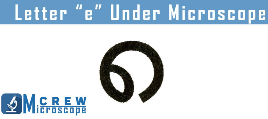
How does the Letter ‘E’ Look Under a Microscope?
The letter ‘e’ appears backward and upside down under a microscope at 40x magnification; you can hardly see the full letter ‘e’ at 100x.
The observation shows the rough edges of the specimen, and you can also observe the texture of the paper in the eyepiece. The ink also looks like it is broken in pieces and not solid.
An increase in magnification also affects how the letter ‘e’ looks under a microscope. You can see the letter ‘e’ 100% at lower magnification, that comes down to 70% on medium magnification. Using a higher magnification of 100x or more massively reduces the field of view, letting you see only 1-10% of the specimen.

Why does the Letter ‘E’ Appear Inverted Under a Microscope?
The letter ‘e’ appears inverted and backward under a microscope due to two sets of lenses in the microscope. These lenses act like mirrors, thus inverting the image while magnifying it.
Besides being inverted, it is also backward. It means that the specimen will move in a direction opposite than intended. So, if you want to observe the slide on the right side, move it to the left.
Depending on your microscope type, some observers also suggest a better way to check the specimen from the other side. You do not have to worry about an inverted or backward image.
At the same time, a few others believe that the brain fixes the image itself if you keep looking at it for a long time, as the retina inverts all the images we see.
However, besides all these theories, the fact is that the letter ‘e’ looks inverted and backward because of the action of two different lenses.
The Bottom Line
While the letter ‘e’ is not one of the most common specimens to observe in a lab, learning how the letter ‘e’ looks under a microscope is an interesting experience. You can observe the letter ‘e’ from a newspaper cutout by preparing a wet mount; it allows you to see the specimen more clearly. Opposed to other samples under a microscope, the letter ‘e’ looks inverted and backward; see how you place the slide under the microscope and how it appears in the eyepiece. Similarly, it is also backward. Move the stage in the opposite direction you want to move the slide in.
FAQs
Why is it necessary to center the letter “e” in the microscope field of view before switching the objective to medium or high power?
Shifting from a low-magnification lens to a high-magnification lens decreases the field of view, making it difficult to find the specimen on the slide. Thus, adjusting the letter ‘e’ in the microscope in the center is recommended before moving to higher magnification.
What happened to the letter E when you moved the slide to the left?
As the letter ‘e’ seems backward under a microscope, it moves in another direction. So, when you move the slide to the left, the letter ‘e’ will move to the right.
Why does the E disappear at high magnification?
A higher magnification means zooming in on the specimen. So, when you increase the magnification of the lens when observing the letter ‘e,’ it reduces the field of view, and you cannot see the full letter under the microscope.
How many times is the image of e magnified when viewed through the high power objective?
You can observe the letter ‘e’ in less than 40x magnification on a standard compound microscope, where you can see the specimen under the lens. Furthermore, viewing it at 100x and 400x is also possible. However, using such high magnification might distort the image, and you will see only a small part of the slide.
Leave a Reply