The human body is an intriguing yet beautiful creation to be absorbed by a common person. It has a list of complex systems functioning side-by-side to make a human body work and grow. Among them are the circulatory, respiratory, lymphatic, nervous, and integumentary systems.
Now if we talk more specifically about the integumentary system, it is the one comprising the largest organ of a body called the skin. Besides the skin are numerous appendages that function together with the skin to protect the body from the external environment. Even though the skin is a flexible physical barrier working at its best as a first-line defense, it still gets abrasions and wounds sometimes.
When a kid or even an adult gets a wound, bacteria attack that open wound and start increasing in number to make biofilms. Now you must be wondering what a wound looks like under a microscope, but worry not! We are here to give you detailed knowledge about wounds and their appearance under a handy microscope.
What are Wounds?
Every one of you will definitely experience getting open wounds at a certain stage of life. Usually, children the age of playing outdoor games fall and encounter such injuries, but what actually are wounds?
In simpler words, wounds are acute or chronic injuries on the skin. When a person gets these scratches due to any reason, it breaks the skin and its nearby tissues. If these lesions stay exposed to the outside environment and do not dissolve within four to eight weeks, they turn into chronic wounds. One should avoid reaching this state as it can lead to amputations in the worst cases.
Types of Wounds
Wounds can be classified into several types based on different metrics, such as the healing time or contamination. These are briefly explained below:
Based on the Surface Type
Open Wounds: If you see an exposed lesion or damaged tissues on the skin, these are called open wounds. You can observe an open wound in its fresh or healing state easily under different kinds of microscopes. There are typically four types of open wounds, including abrasions, punctures, lacerations, and avulsions.
Closed Wound: Unlike an open wound, a closed one is a non-penetrating injury that has internal damage to the body. There is no exposure of the underlying damaged tissues or organs and such wounds usually occur straight due to a trauma or severe injury. Common types of closed wounds are blisters, contusions, hematomas, and seromas.
Based on Contamination
Clean Wounds: As the name suggests, clean wounds are those with zero inflammation and no signs of growing infection. These wounds heal naturally and cause no damage to the internal organ system. Clean wounds usually appear in the eyes and the vascular system.
Contaminated Wounds: As the name indicates, these are fresh wounds covered with biofilms of bacteria and can lead to severe infections. When a person experiences any injury, cut, or scratch, it breaks the skin to create a pace, and the bacteria multiplies, making it contaminated. When a fresh wound starts healing with time, it is called a healing wound.
Do you wonder how do these two types look like under a simple microscope? Let’s explore with us now.
Fresh Wound under a Microscope
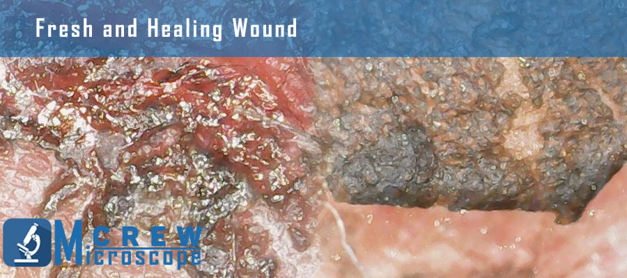
When an exposed wound, on any body part, is in the starting or initial phase, it is called a fresh wound. Below is a picture of a fresh wound on the knee of a preschooler. It is taken after observing it under a microscope with 50X magnification.
Healing Wound under a Microscope
When the wound has passed almost four weeks and starts proliferating after going through inflammation, the state is called a healing wound. As you can see in the above picture, there is a representation of a healing wound besides a fresh wound. Both these pictures are taken from a handy microscope with a maximum magnification of 50X.
Microscopic Video of a Wound
Just in case you’ve missed understanding how a wound look under a microscope from the above pictures, here is a video of it as well:
Conclusion
Different types and even different states of wounds can be observed under a microscope. You can choose to see it under a simple light microscope, or compound microscope, and more details can be seen under a digital microscope. However, we have given details of fresh and healing wounds under a microscope having 50X as maximum magnification. We hope to clarify the basics and your perception of how it appears under a microscope.
Leave a Reply